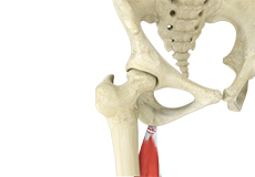
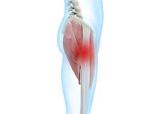
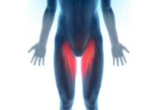
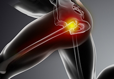
Hamstring Injuries
Hamstring injuries occur when these muscles are strained or pulled. They are common in dancers and athletes of all sorts including runners and those who play football, soccer, basketball, tennis, etc.
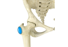
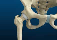
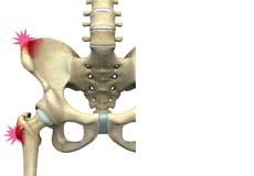
Hip Bursitis
Hip bursitis is a painful condition caused by the inflammation of a bursa in the hip. Bursae are fluid-filled sacs present in the joints between bone and soft tissue to reduce friction and provide cushioning during movement.
Gluteus Tendon Tear
The gluteal muscles (situated in the buttocks) are necessary for the stability and movement of the hip joints. The tendons of two gluteal muscles (gluteus medius and gluteal minimus) are attached at the outer hip region and are often called the “rotator cuff of the hip.” These tendons may be subject to injury or tearing due to various reasons. Since these gluteal muscles are involved in abduction (movement of your leg away from the midline of the body), the tears are also called abductor tendon tears.
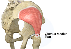
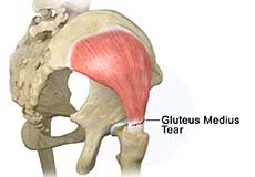
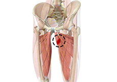
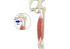
Gluteus Medius Tear
A gluteus medius tear is the partial or complete rupture of the gluteus medius muscle due to severe muscle strain.
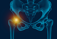
Hip Injury
The hip joint is one of the most important and flexible joints in the human body which allows us to walk, run, bend and perform physical activities.
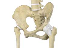
Hip Ligament Injuries
Injuries to the hip ligaments are commonly called a hip sprain and can range from minor tears of the ligaments to more serious injuries involving the hip muscles, tendons or bone.
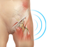
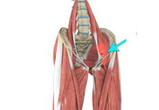
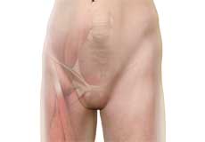
Hip Flexor Pain
Hip flexor pain is a distressing feeling or discomfort noted in the hip and/or groin region that can make everyday activities, such as going up and down the stairs or lifting your leg to tie a shoe extremely difficult and painful and can severely limit your activity and mobility.
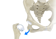
Hip Dislocation
The hip joint is a “ball and socket” joint. The “ball” is the head of the femur or thighbone, and the “socket” is the cup-shaped acetabulum.
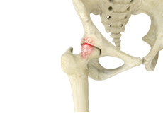
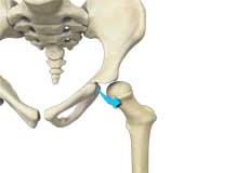
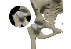
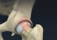
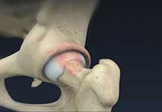
Hip Labral Tear
A hip labral tear is an injury to the labrum, the cartilage that surrounds the outside rim of your hip joint socket.

Hip Instability
Injury or damage to these structures can lead to a condition called hip instability when the joint becomes unstable.
Hip Abductor Tears
Hip abductors are a major group of muscles found in the buttocks. It includes the gluteus maximus, gluteus medius, gluteus minimus, and tensor fascia lata muscles.
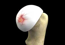
Avascular Necrosis
Bones are living tissue relying on blood vessels to bring blood to keep them alive. Most living tissues have blood vessels that come from many directions into the tissue.
Femoroacetabular Impingement
Femoroacetabular impingement (FAI) is a condition characterized by excessive friction in the hip joint from the presence of bony irregularities.
Snapping Hip Syndrome
Snapping hip syndrome is a condition in which you hear or feel a snapping sound in the hip when you swing your legs, run, walk or get up from a chair. The sound can be experienced in the back, front or side of the hip.
Hip Adductor Injuries
Hip adductors are the group of muscles on the inner side of your thigh that enable adduction or the ability to bring the thighs together.
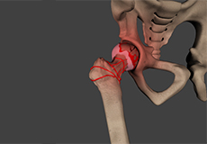
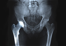
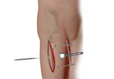
Stress Fractures of the Hip
Stress fractures of the hip are a break in the upper part of the thigh bone (femur) that fits into the socket of the hip joint. It can occur in any part of the hip, however, it mostly occurs just below the ball of the ball-and-socket hip joint called the femoral neck.
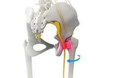
Greater Trochanteric Pain Syndrome
GTPS, also called trochanteric bursitis, is a localized painful condition affecting your outer thigh and hip area where the pain is typically confined to only the outer side of the femur at the edge of the hip.
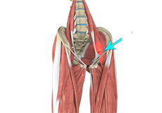
Hip Flexor Strain
A hip flexor strain is an overuse injury to the flexor muscles of your hip and can range from a minor stretch injury to a complete tear of the muscle fibers or tendons.
Iliopsoas Tendonitis
Iliopsoas tendonitis also referred to as snapping hip syndrome, is an inflammation of the iliopsoas tendon or the surrounding area. The iliopsoas is the hip flexor tendon located over the front of the hip socket. The term snapping hip describes the sound made, a snap or click, that occurs with certain hip movements including flexion, extension, and rotation of the hip.
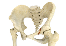
Avulsion Fractures of the Pelvis
Avulsion fractures of the pelvis is an injury that occurs when a tendon or ligament pulls off a piece of bone from the hip.
Borderline Hip Dysplasia
Hip dysplasia is a medical condition where the acetabulum (hip socket) does not fully cover the ball-like head at the top of the femur (thighbone). Most people who have hip dysplasia are born with it.
Hip Groin Disorders
Hip and groin disorders are more common in athletes. They are caused by rapid acceleration and deceleration motion.
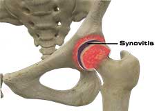
Hip Synovitis
Hip synovitis, also called transient hip synovitis or toxic synovitis, is a condition characterized by inflammation of the synovial tissues that surround the hip joint. It is the most common cause for sudden hip pain that occurs in young children between the age of 2 and 9. It affects boys more commonly than girls and is most of the time limited to only one side of the hip.
Hip Tendonitis
Tendons are strong connective tissue structures that connect muscle to bone. Hip tendonitis is a condition associated with degeneration of the hip tendons. This condition is mainly caused due to strain on the tendons which may occur due to overuse or biomechanical problems.
Hip Pointer
The hip joint consists of the femur (thighbone) and pelvic bone, which is made up of the fusion of three bones – the ischium, pubis, and ilium. The femur has two boney prominences close to the hip joint – the greater and lesser trochanters. Hip pointer is an injury or bruise to the iliac crest (curved upper border of the ilium) or greater trochanter, or the surrounding muscles or tissues.
Transient Osteoporosis of the Hip
Transient osteoporosis of the hip is a rare condition that causes temporary bone loss in the upper region of the thighbone (femur). It occurs most often in young or middle-aged men of the age groups 30 to 60, and women in their later stages of pregnancy or early postpartum (following childbirth). It is characterized by abrupt onset of pain that increases with activity.
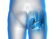
Groin Injuries in Athletes
Groin injuries are injuries sustained by athletes during sports activity. Groin injuries comprise about 2 to 5 percent of all sports injuries. The most common kind of groin injury is a groin strain or a pulled groin muscle. Typical groin injuries vary based on athlete’s gender, age, and sports, with most common groin injuries occurring in certain sports such as football with a prevalence rate as high as 12-16%.
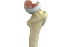
Hip Osteonecrosis
Hip osteonecrosis occurs due to disruption of the blood supply to the highest part of the thigh bone (femoral head). Due to lack of nourishment, the bone tissue of the femoral head dies and gradually collapses, which may further lead to degeneration of the underlying cartilage.
Hip Preservation Surgery
The hip is a ball and socket joint comprising of the femur (thigh bone) and the pelvic bone. The head of the femur (ball) articulates with a cavity (socket) called the acetabulum in the pelvic bone. To facilitate the smooth and frictionless movement of the hip joint, the articulating surfaces of the femur head and acetabulum are covered by spongy articular cartilage.
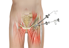
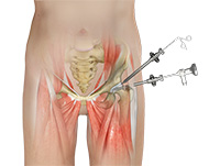
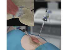
Hip Arthroscopy
Hip arthroscopy, also referred to as keyhole or minimally invasive surgery, is a procedure in which an arthroscope is inserted into your hip joint to check for any damage and repair it simultaneously.
Hip Cartilage Repair
Hip cartilage is a white, tough, flexible tissue covering the ball (femoral head) and socket (acetabulum) of your hip joint. It acts as a cushion or shock-absorber and allows the bones to slide over one another by providing a smooth surface in the joint.
Hip Labral Repair
Labrum is a ring of strong fibrocartilaginous tissue lining around the socket of the hip joint. Labrum serves many functions where it acts as a shock absorber, lubricates the joint, and distributes the pressure equally. It holds the head of the femur in place and prevents the lateral and vertical movement of the femur head within the joint. It also deepens the acetabular cavity and offers stability against femoral head translation.
Hip Resurfacing
The hip joint is also known as a ball and socket joint, where the ball (femoral head) of the thigh bone fits into the socket (acetabulum) of the pelvic bone.
Proximal Hamstring Repair
These muscles help in extending your leg and bending your knee. Therefore, any damage to the hamstring muscle group affects both hip and knee movements.
Hip Cartilage Restoration
Hip cartilage restoration is a surgical technique to repair damaged articular cartilage in the hip joint by stimulating new growth of cartilage or by transplanting cartilage into areas with defects in order to relieve pain and restore normal function to the hip.
Complex Hip Reconstruction Surgery
Complex hip reconstruction surgery is a surgical procedure employed to treat hip structures with complex hip fractures or traumatic hip injuries, deformities, structural issues, and damage from diseases such as arthritis. The main objective of complex hip reconstruction surgery is to alleviate hip pain and stiffness, improve range of motion, and restore normal functioning of the hip joint to help you resume your normal activities and improve your quality of life.
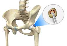
Hip Microfracture
Hip Microfracture is a marrow-stimulating technique that creates a network of small holes in the bone below the hip cartilage (subchondral bone). The goal of the procedure is to increase the blood supply to stimulate cartilage growth.
Hip Capsular Closure
The hip capsule is a membranous fold consisting of three major ligaments enveloping the hip joint. It protects the joint and stabilizes it by keeping the joint in position.
Femoral Osteoplasty
Osteoplasty is a surgery performed to repair and redesign a bone to its original shape. The surgery is used to treat bone deformation in various joints of the body.

Endoscopic Surgery for Deep Gluteal Syndrome
Deep gluteal syndrome (DGS) is an underdiagnosed medical condition characterized by symptoms of excruciating pain, numbness, and tingling in the buttock, hip, or posterior thigh area that can radiate down the back of the leg. The symptoms are caused by compression of the sciatic nerve in the subgluteal space by the piriformis muscle in the buttock area due to muscular hypertrophy, trauma to the hip or pelvis, or anatomical abnormalities.
Core Decompression for Avascular Necrosis of the Hip
The hip joint is a ball and socket joint, where the head of the thighbone (femur) articulates with the cavity (acetabulum) of the pelvic bone.
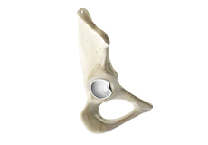
Periacetabular Osteotomy
Periacetabular osteotomy is a surgical procedure to treat a congenital hip condition called hip dysplasia. Hip dysplasia is either present from birth or develops in the first few months of life. Patients suffering from this condition have a shallow socket (acetabulum) of the hip joint. This causes misalignment of the head of the thigh bone (femur) in the acetabulum and leads to premature wear and tear of the joint over the years.
Postless Hip Arthroscopy
The hip joint is a ball and socket joint and is surrounded by muscles, ligaments, and tendons. Traditionally, access to the hip joint for surgery was achieved by placing traction to the operative leg and perineal posts (wide-diameter post) in the groin region.
Arthroscopic Gluteus Medius Tendon Repair
Arthroscopic gluteus medius tendon repair is a minimally invasive surgical procedure employed for the treatment of a gluteus medius tendon tear, when the tear does not respond to conservative treatment.

Acetabular Rim Trimming
Acetabular rim trimming is a surgical procedure performed to even the surface around the femoral head (the topmost portion of the thigh bone) of your hip joint.
Femoroacetabular Osteoplasty
Femoroacetabular Osteoplasty is a surgical procedure performed to even the surface around the femoral head (the topmost portion of the thigh bone) of your hip joint.
Ischiofemoral Impingement Decompression - Procedure
Ischiofemoral impingement decompression is a surgery performed to relieve hip pain caused by entrapment or compression of soft tissue in the ischiofermoral space present between the upper part of the thigh bone and the lower back part of the hip bone.
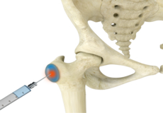
Trochanteric Bursa Injections
A trochanteric bursa injection is a minimally invasive procedure in which medicine is injected directly into the trochanteric bursa in the hip joint using a thin needle and syringe to relieve pain and inflammation. The injection usually contains a combination of numbing medicine and cortisone (an anti-inflammatory agent). Trochanteric bursitis, also known as greater trochanteric bursitis or hip bursitis, is the main indication for a trochanteric bursa injection.
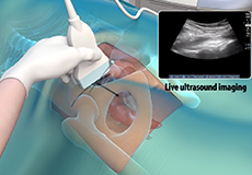
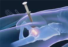
Ultrasound Guided Hip Injections
An ultrasound scan is an imaging procedure that uses high-frequency sound waves to produce pictures of the inside of the body. Ultrasound-guided hip joint injections are used to diagnose the underlying cause and relieve hip pain. The injection consists of a special mixture of an anesthetic and a steroid that blocks pain impulses and reduces inflammation in the injected area.
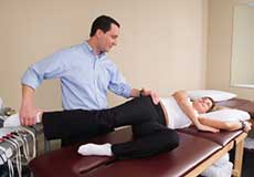
Physical Therapy for Hip
Physical therapy is an exercise program that helps you to improve movement, relieve pain, encourage blood flow for faster healing, and restore your physical function and fitness level.
Stem Cell Therapy for Hip Injuries
Stem cell therapy is a form of regenerative medicine that utilizes the body’s natural healing mechanism to treat various conditions.
Physical Examination of the Hip
The physical examination of the hip by your doctor includes a visual inspection of your hip, palpation of the hip to diagnose tenderness or any abnormality, etc; and testing range of motion of the hip.
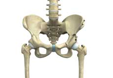
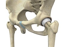
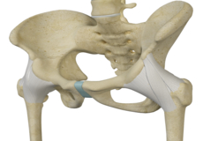
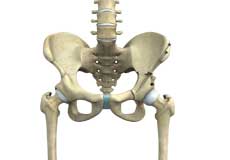
Hip Joint
The hip joint is the largest weight-bearing joint in the human body. It is also referred to as a ball and socket joint and is surrounded by muscles, ligaments, and tendons. The thigh bone or femur and the pelvis join to form the hip joint.
Any injury or disease of the hip will adversely affect the joint's range of motion and ability to bear weight.
The hip joint is made up of the following:
- Bones and joints
- Ligaments of the joint capsule
- Muscles and tendons
- Nerves and blood vessels that supply the bones and muscles of the hip
Bones and Joints
The hip joint is the junction where the hip joins the leg to the trunk of the body. It is comprised of two bones: the thigh bone or femur and the pelvis which is made up of three bones called ilium, ischium, and pubis. The ball of the hip joint is made by the femoral head while the socket is formed by the acetabulum. The Acetabulum is a deep, circular socket formed on the outer edge of the pelvis by the union of three bones: ilium, ischium, and pubis. The lower part of the ilium is attached by the pubis while the ischium is considerably behind the pubis. The stability of the hip is provided by the joint capsule or acetabulum and the muscles and ligaments which surround and support the hip joint.
The head of the femur rotates and glides within the acetabulum. A fibrocartilagenous lining called the labrum is attached to the acetabulum and further increases the depth of the socket.
The femur or thigh bone is one of the longest bones in the human body. The upper part of the thigh bone consists of the femoral head, femoral neck, and greater and lesser trochanters. The head of the femur joins the pelvis (acetabulum) to form the hip joint. Next, to the femoral neck, there are two protrusions known as greater and lesser trochanters which serve as sites of muscle attachment.
Articular cartilage is the thin, tough, flexible, and slippery surface lubricated by synovial fluid that covers the weight-bearing bones of the body. It enables smooth movements of the bones and reduces friction.
Ligaments
Ligaments are fibrous structures that connect bones to other bones. The hip joint is encircled with ligaments to provide stability to the hip by forming a dense and fibrous structure around the joint capsule. The ligaments adjoining the hip joint include:
- Iliofemoral ligament: This is a Y-shaped ligament that connects the pelvis to the femoral head at the front of the joint. It helps in limiting the over-extension of the hip.
- Pubofemoral ligament: This is a triangular shaped ligament that extends between the upper portion of the pubis and the iliofemoral ligament. It attaches the pubis to the femoral head.
- Ischiofemoral ligament: This is a group of strong fibers that arise from the ischium behind the acetabulum and merge with the fibers of the joint capsule.
- Ligamentum teres: This is a small ligament that extends from the tip of the femoral head to the acetabulum. Although it has no role in hip movement, it does have a small artery within that supplies blood to a part of the femoral head.
- Acetabular labrum: The labrum is a fibrous cartilage ring which lines the acetabular socket. It deepens the cavity, increasing the stability and strength of the hip joint.
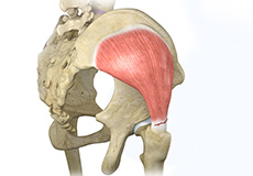
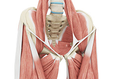
Muscles and Tendons
A long tendon called the iliotibial band runs along the femur from the hip to the knee and serves as an attachment site for several hip muscles including the following:
- Gluteals: These are the muscles that form the buttocks. There are three muscles (gluteus minimus, gluteus maximus, and gluteus medius) that attach to the back of the pelvis and insert into the greater trochanter of the femur.
- Adductors: These muscles are located in the thigh which helps in adduction, the action of pulling the leg back towards the midline.
- Iliopsoas: This muscle is located in front of the hip joint and provides flexion. It is a deep muscle that originates from the lower back and pelvis and extends up to the inside surface of the upper part of the femur.
- Rectus femoris: This is the largest band of muscles located in front of the thigh. They also are hip flexors.
- Hamstring muscles: These begin at the bottom of the pelvis and run down the back of the thigh. Because they cross the back of the hip joint, they help in extension of the hip by pulling it backward.
Nerves and Arteries
Nerves of the hip transfer signals from the brain to the muscles to aid in hip movement. They also carry the sensory signals such as touch, pain, and temperature back to the brain.
The main nerves in the hip region include the femoral nerve in the front of the femur and the sciatic nerve at the back. The hip is also supplied by a smaller nerve known as the obturator nerve.
In addition to these nerves, there are blood vessels that supply blood to the lower limbs. The femoral artery, one of the largest arteries in the body, arises deep in the pelvis and can be felt in front of the upper thigh.
Hip Movements
All of the anatomical parts of the hip work together to enable various hip movements. Hip movements include flexion, extension, abduction, adduction, circumduction, and hip rotation.